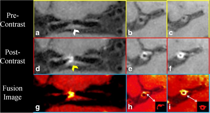
Fig. 1.
Pre-contrast coronal and cross-sectional whole brain high resolution cardiovascular magnetic resonance (WB-HRCMR) images showed diffused plaque (a-c, white arrow) located on middle cerebral artery (MCA). Partial enhancement of the plaque was observed (d-f, yellow arrow). The enhancement plaque area was segmented through fusion images (h, i). The plaque enhanced volume was 11.02mm3

