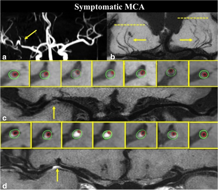
Fig. 3.
A 61 years old symptomatic ICAS patient with severe stenosis on right MCA (a), coronal MinIP revealed the decrease of right LSA branches compared to the left side (b); pre-contrast curved WB-HRCMR and cross-sectional images showed a plaque (c, arrow) on the MCA wall; Post-contrast WB-HRCMR showed extensive enhanced plaque volume which can be measured on corresponding cross-sectional images

