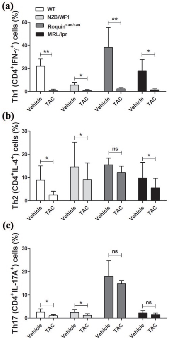
Figure 1.

Suppression of effector T cells by tacrolimus (TAC) in mice. Splenocytes from the spleen of wild-type (WT) or lupus-prone NZB/WF1, Roquinsan/san, and MRL/lpr mice (n = 5) were stimulated with TAC (1 nM) in the presence of anti-CD3 and anti-CD28 for 3 days. Cells were stimulated with phorbol myristate acetate (PMA), ionomycin, and GolgiStop for 4 h and stained with antibodies against (a) CD4+IFN-γ+ Th1 cells, (b) CD4+IL-4+ Th2 cells, and (c) CD4+IL-17A+ Th17 cells for intracellular flow cytometric analysis. *P < 0.05, **P < 0.01 versus vehicle-treated condition. Data are mean ± standard deviation (SD).
