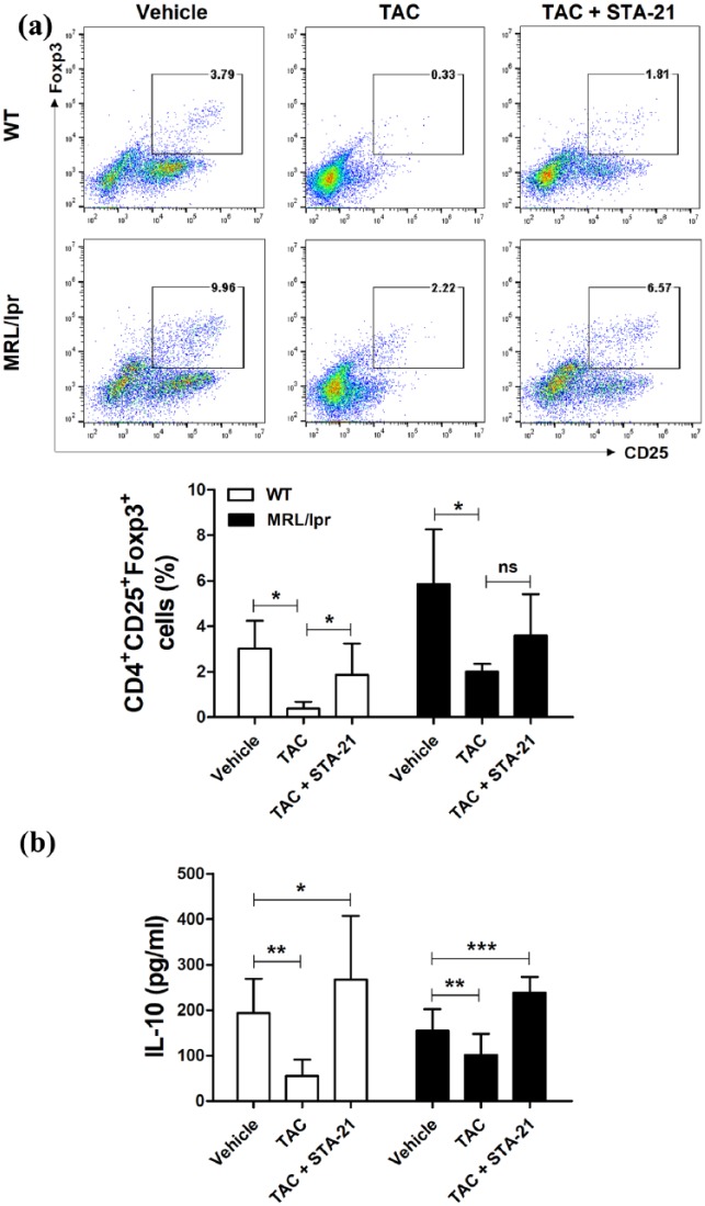
Figure 3.
TAC and STA-21 induced Treg cells. Splenocytes of WT or lupus-prone MRL/lpr mice (n = 7) were cultured with TAC (1 nM) and STA-21 (10 μM) in the presence of anti-CD3 and anti-CD28 for 3 days. (a) After 3 days, cells were stained with antibodies against CD4+CD25+Foxp3+ Treg cells for intracellular flow cytometric analysis. (b) IL-10 concentrations in culture supernatants were determined by ELISA. *P < 0.05, **P < 0.01, ***P < 0.001. Data are mean ± SD.

