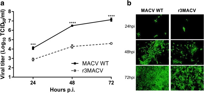
Fig. 2.
Growth properties of the r3MACV. VERO cells were infected for one hour with the MACV wt or the r3MACV viruses at a MOI of 0.01. The inoculum was then removed and with culture medium (DMEM GlutaMax 1% supplemented with 2% fetal bovine serum) was added. a At indicated time points, virus titers in the supernatant solutions were determined by virus TCID50 (50% tissue culture infective dose) using the end-point dilution assay and the Reed-Müench calculation method. Viral titers were determined in octuplicates using immunofluorescence [11] with the primary mouse monoclonal antibody anti-MACV NP. b At indicated time points, cells were fixed with PFA 4% (Electron microscopy sciences) and permeabilized with PBS containing 0.3% Triton X-100 (Merck) and 3% bovine serum albumin (Sigma). Viral NP expression was revealed using an Alexa Fluor 488 secondary antibody (Thermo Fisher). An epifluorescence optical microscope (× 100) was used for the eGFP observation. This experiment was done in triplicate. Asterisks denote significant differences (P < 0.05, two-way ANOVA with the Bonferroni correction, *** ≤ 0.001, **** ≤ 0.0001)

