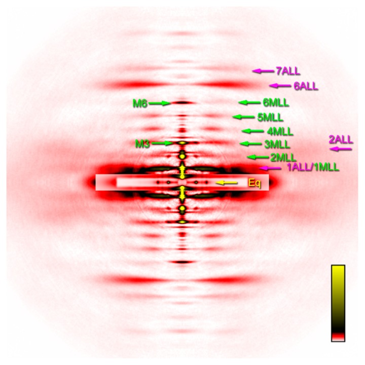
Figure 4.

Two-dimensional diffraction pattern summed from 4 sets of 30 muscle fibers. MLL, myosin layer-line reflection; ALL, actin layer-line reflection. The number is the order of reflection. The 1st actin layer-line reflection at 1/36 nm and the 1st myosin reflection at 1/42.9 nm are partially overlapped. Eq, equatorial reflections. Of the two prominent reflections, the inner one is the 1,0 and the outer one is the 1,1. M3 and M6 are the myosin meridional reflections at 1/14.3 nm and 1/7.2 nm, respectively. The pattern was recorded in the presence of 100 μM blebbistatin at pCa=4.0 at 6–8°C.
