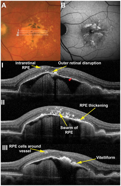
Figure 4. Histologically defined features are visible in vivo.
A large drusenoid pigment epithelial detachment (D-PED) in a 77 year-old male demonstrates pigmentary changes on the surface that correlate to sites of increased fundus autofluorescence (B). Areas of B-scans (I, II and III) are illustrated in the color image. A range of RPE related changes are seen in OCT scans including intraretinal RPE cells and vitelliform lesions. Note that the ellipsoid zone (red arrowhead) is visible on the surface of the PED with the exception of the apex where it is notably absent. In this case it was possible to distinguish vitelliform lesions from RPE thickening however this distinction was not possible in most cases in this series.

