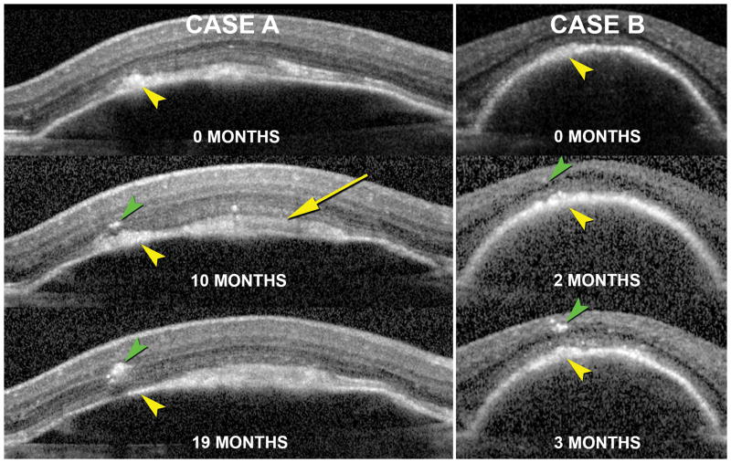
Figure 5. Association of intraretinal hyperreflective foci and thickening of the RPE+BL band during the lifecycle of drusenoid PEDs (D-PED).
The natural course of D-PED of a 71 year-old female (Case A) and 68 year-old female (Case B) as seen using in vivo SD-OCT with image registration enabled are presented. At their first visit (0 months) hyperreflective material is seen at the level of the RPE and subretinal space (yellow arrows) in both cases. Over the course of time, hyperreflective foci are seen to migrate from the RPE+BL band into the retina (green arrowheads). The rate of RPE migration and the quantity of cells migrating into the retina were different in the two cases. Yellow arrowheads denote the same location in the PED in image-registered sections and may signify vitelliform material in addition to RPE cell bodies in transit. White arrow (Case A, 10 month) also shows vitelliform material.

