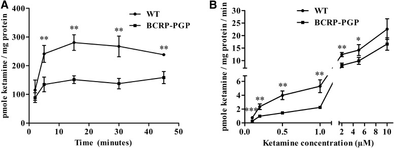
Fig. 4.
3H-ketamine uptake is lower in MDCKII BCRP-PGP cells compared with MDCKII WT cells. (A) Time-dependent uptake of 3H-ketamine in WT and BCRP-PGP transfected MDCKII cells. Ketamine’s intracellular concentration at each time point was compared between the WT and BCRP-PGP cells using an unpaired t test. **P < 0.01. (B) 3H-ketamine uptake in WT and BCRP-PGP transfected MDCKII cells at different ketamine concentrations. The intracellular concentration of 3H-ketamine at each treatment was compared between the WT and BCRP-PGP cells using an unpaired t test. *P < 0.05; **P < 0.01; ***P < 0.001.

