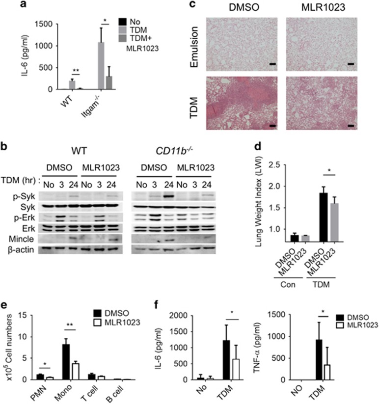
Figure 9.
The Lyn activator MLR1023 inhibits TDM signaling both in vivo and in vitro. (a) Production of IL-6 by WT and CD11b−/− BMMs challenged with TDM and MLR1023 (1 ng ml−1) or DMSO for 24 h was assessed by ELISA. (b) Western blotting assay of Syk and Erk kinase activity and Mincle induction in WT and CD11b−/− BMMs treated with TDM and MLR1023 (1 ng ml−1) or DMSO for 24 h. β-Actin protein expression was used as a loading control. Experimental and control mice (n=8 mice per group) were intravenously injected with 3.75 mg kg−1 TDM in an oil-in-water emulsion on day 0. Then the experimental mice were intraperitoneally injected with MLR1023 (6 mg kg−1 in PBS) every day beginning on day 1, until the mice were killed 7 days post-TDM challenge. The control mice were injected with 1% DMSO in PBS. (c) Lung tissues were isolated and stained with hematoxylin and eosin (H&E) for histology analysis after the lung weight index (LWI) (d) was determined. (e) Leukocyte subsets were analyzed by flow cytometry using distinct markers for monocytes and macrophages (Mo/Ma; CD11b+ Ly6G−), neutrophils (PMN; CD11b+ Ly6G+), T cells (CD3+) and B cells (CD19+). (f) Lung homogenates were analyzed by ELISA for TNF-α and IL-6. Data are representative of two independent experiments. (c–f, mean and s.d. of eight mice per group.) *P<0.05, **P<0.01 (two-tailed unpaired Student’s t-test).

