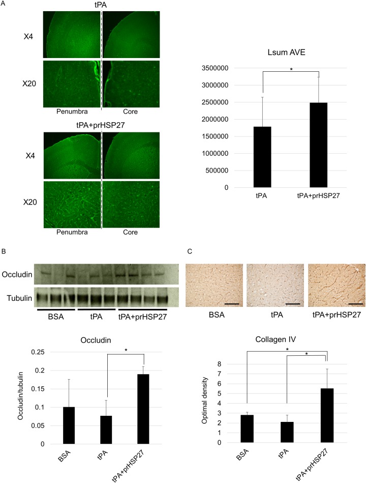
Fig 4. prHSP27 maintains the cell wall structure of endothelial cells and tight junction proteins.
(A) Localization of endothelial cell-stained FITC-tomato lectin on the penumbra and core of the ischemic side of the brain from mice treated with tPA or tPA plus prHSP27. FITC-staining is higher in the mice treated with prHSP27. *p < 0.05: tPA vs tPA plus prHSP27. (B) Immunoblot analysis (upper) and quantitation (lower) of occludin. Protein loading was calculated relative to tubulin. ‡p < 0.005: tPA plus prHSP27 vs. tPA. (C) Photomicrographs (upper) and quantitation (lower) of type IV collagen immunostaining in the infarct boundary zones in BSA- (n = 5), tPA- (n = 5), and tPA plus prHSP27- (n = 5) treated mice at 24 h after reperfusion. prHSP27 maintained the type IV collagen structure of the basement membrane. Scale bars = 100 μm. Data are mean ± SEM. *p < 0.05: tPA plus prHSP27 vs. BSA, tPA plus prHSP27 vs. tPA.

