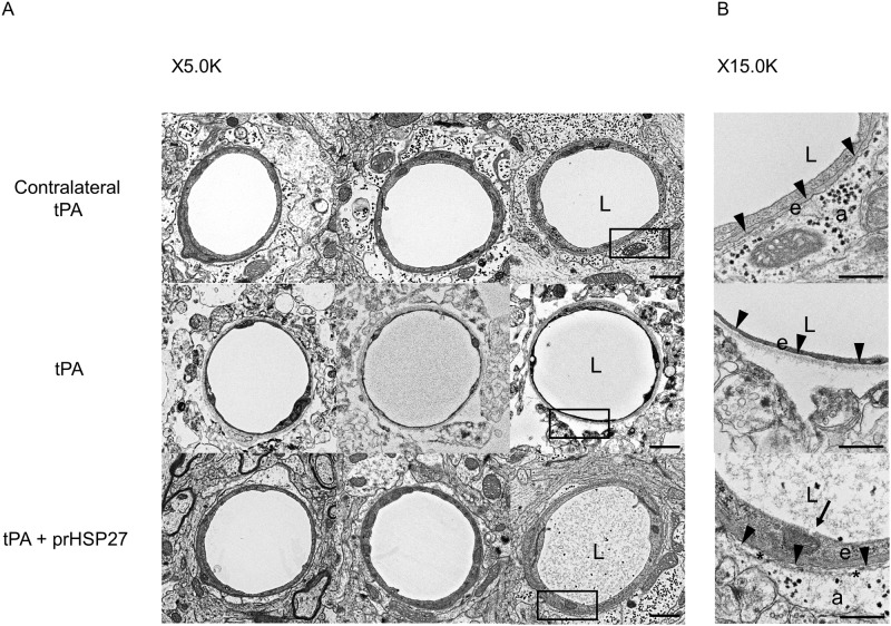
Fig 6. Effect of cerebral ischemia on prHSP27 in the blood-brain barrier.
(A) Electron microscopic analysis of the blood-brain barrier on the non-ischemic side of tPA- (contralateral tPA, n = 3), ischemic side of tPA- (tPA, n = 3), and ischemic side of tPA plus prHSP27- (tPA and prHSP27, n = 3) treated mice. Magnification, ×5000 (left row) and ×15000 (right row). The left row shows magnifications of the boxed areas in the right row. Scale bars, 1 μm (left row) and 500 nm (right row). a: astrocyte endfeet; e: endothelial cell; L: lumen. Arrowheads: the space between the basement membrane and astrocyte endfeet. Asterisks indicate the space between the basement membrane and astrocyte endfeet (B).

