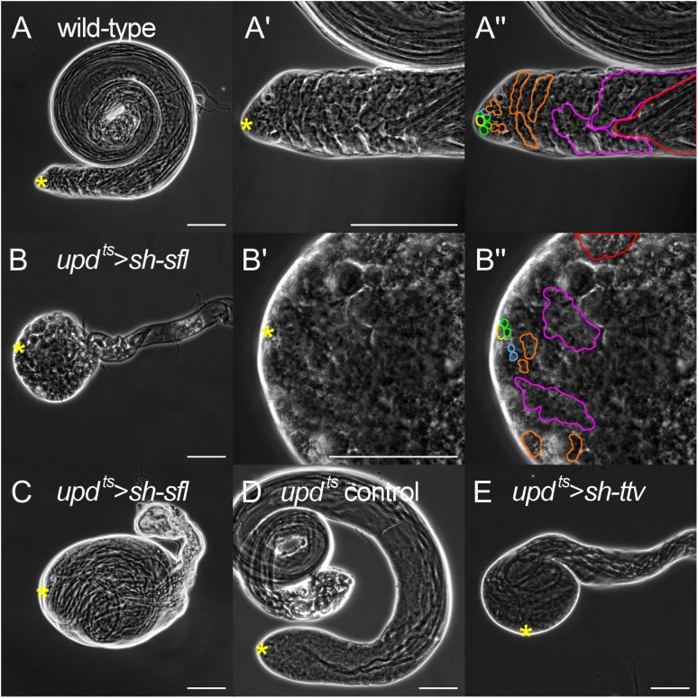
Fig. 1.
Loss of hub HS results in tumorous testes and disrupts organization of spermatogenic cells. Phase contrast images of testes from wild-type (A–A”), updts > sh-sfl (B–C), updts control (D) and updts > sh-ttv (E). A’–A” and B’–B” are high magnification views of A and B, respectively, showing organization of spermatogenic cells. To illustrate the progressive stages of differentiation as cells transit the testis, a few clearly identified examples of germline cell clusters at different stages of spermatogenesis are highlighted by outline color in A” and B”: yellow, hub; green, GSCs; blue, gonialblasts; orange, spermatogonia; pink, primary spermatocytes; red, elongating spermatids. The cells in wild-type advance through each stage of spermatogenesis in a successive, linear fashion from the testis tip distally. In updts > sh-sfl testes, however, at least a portion of cells do not follow this normal, linear organization of differentiating cells. Severe “tumorous” phenotypes are seen in hub sfl RNAi (B and C) and hub ttv RNAi (E) testes. Yellow asterisks indicate hub location. Bars: 100 μm. This figure is available in black and white in print and in color at Glycobiologyonline.

