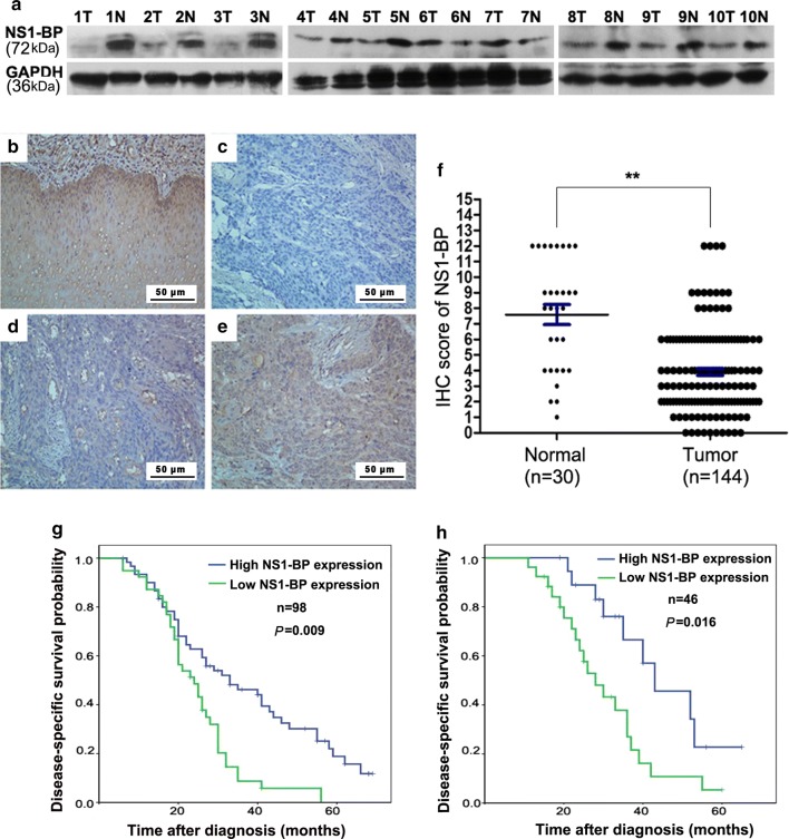
Fig. 1.
NS1-BP expression in esophageal squamous cell carcinoma (ESCC) and its prognostic significance in ESCC patients. a Western blot showing lower NS1-BP levels in ESCC tissues than adjacent non-neoplastic esophageal mucosa tissues (ANT) in 8 of 10 cases. b Normal esophageal mucosa specimen showing strong staining of NS1-BP (IHC score = 12). c ESCC sample (case 15) exhibiting negative NS1-BP staining. d ESCC sample (case 8) exhibiting low NS1-BP expression (IHC score = 3). e ESCC sample (case 60) exhibiting high NS1-BP expression (IHC score = 9). f Statistical analysis of significantly low expression of NS1-BP in ESCC tissues (**P < 0.01). g, h Low expression of NS1-BP was associated with poor prognosis of ESCC patients. Kaplan–Meier plots showing disease-specific survival in 98 ESCC patients in the training cohort (g) and 46 ESCC patients in the validation cohort (h), according to NS1-BP expression levels in the primary tumor (P = 0.009 and 0.016, log-rank test)

