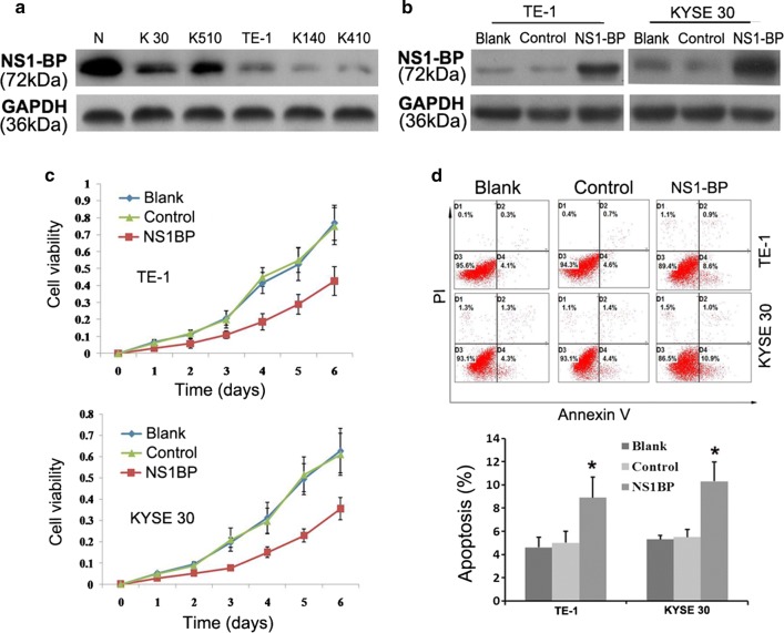
Fig. 2.
Effect of NS1-BP overexpression on ESCC cell proliferation and apoptosis. a Western blot showing that the levels of NS1-BP in five ESCC cell lines (KYSE-30, KYSE-510, TE-1, KYSE-140, and KYSE-410) were lower than in control normal esophageal cells (N). b Western blot showing the expression of NS1-BP in stable transfected TE-1 and KYSE-30 cells (TE-1-NS1-BP; KYSE-30-NS1-BP) relative to empty vector control cells (TE-1-Vector; KYSE-30-Vector) and blank control cells (TE-1-Blank; KYSE-30-Blank). Expression was normalized against endogenous GAPDH. c Cell growth rate was decreased by ectopic overexpression of NS-BP in TE-1 and KYSE-30 cells, as detected by the 3-(4,5-dimethylthiazol-2-yl)-2,5-diphenyl tetrazolium bromide assay. Results are expressed as mean ± standard deviation (SD) of three independent experiments. d NS1-BP promoted tumor cell apoptosis in TE-1 and KYSE-30 cells compared with vector-transfected control cells and blank control cells under normal conditions. Cell apoptotic death events were monitored by annexin V/propidium iodide (PI) staining and flow cytometry assays. The percentage of apoptotic cells is shown as the mean ± SD of three independent experiments. Data represent mean values and SD (*P < 0.05, Student’s t test)

