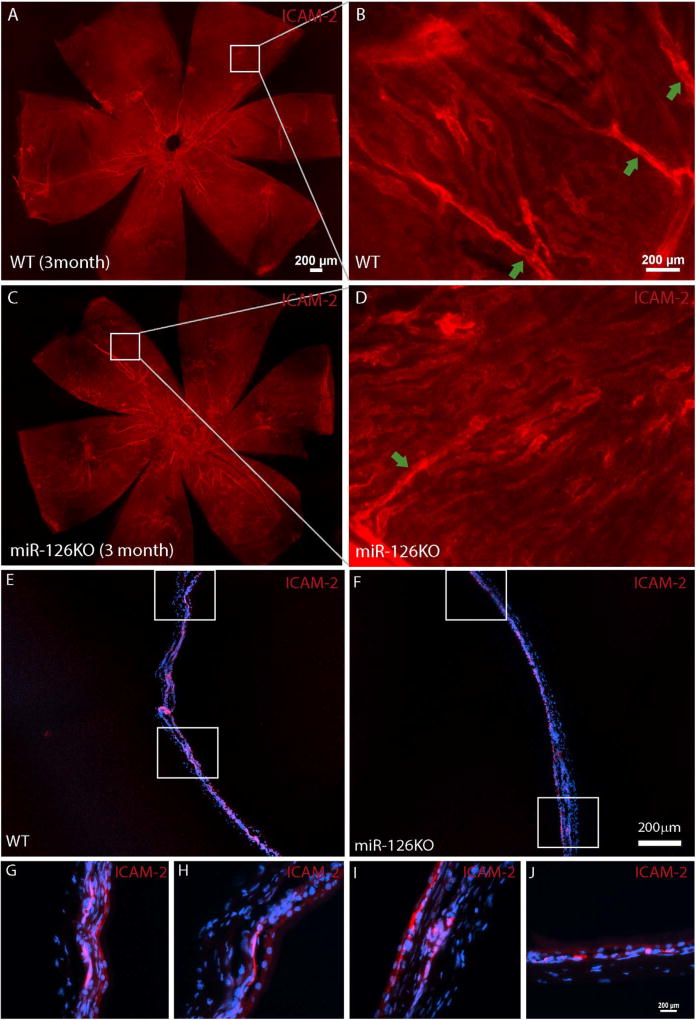
Fig. 4. Less choroid branches but largely normal choroidal vasculature in adult miR-126−/− mice.
(A) Flatmount ICAM-2 staining of a 3 month WT choroid; (B) Magnified picture of the boxed region in (A) showing several vascular branches connecting to the choroidal vascular bed; (C) Flatmount ICAM-2 staining of a 3 month miR-126−/− choroid; (D) Magnified picture of the boxed region in (C) showing one vascular branch connecting to the choroidal vascular bed; (E) Frozen section of the sample in (A); (F) Frozen section of the sample in (B); (G–H) Boxed regions in (A); (I–J) Boxed regions in (B). Scale bar equals 200µm.

