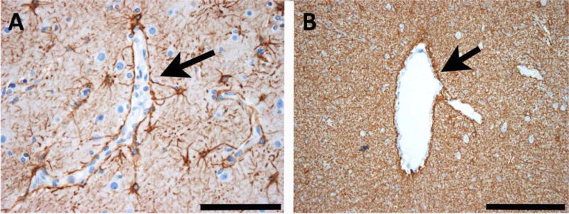
Fig. 2. Common patterns of astrogliosis in epileptic human brain tissue.

A: Reactive astrogliosis in the neocortex of human epilepsy surgery brain specimens is commonly seen along cortical capillaries (arrow). B: In white matter, there is another common pattern of astrogliosis built by a dense glial fibrillary meshwork. The arrow points towards an enlarged venous vessel. Scale bar in A = 100 μm, in B = 200 μm. GFAP immunohistochemistry (brownish color) with bluish hematoxylin counterstaining.
