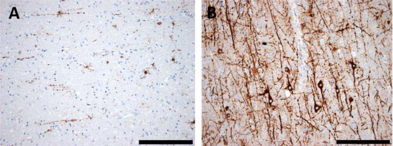
Fig. 3. Common patterns of heterotopic neurons in the white matter of epileptic human brain tissue.

A: The white matter of human temporal lobe usually contains only single heterotopic neurons. B: In many epilepsy specimens of human temporal lobe, there is a vast excess of heterotopic neurons with ramifying neuronal processes in white matter. Scale bar in A and B = 200 μm. MAP2 immunohistochemistry (brownish color) with bluish hematoxylin counterstaining.
