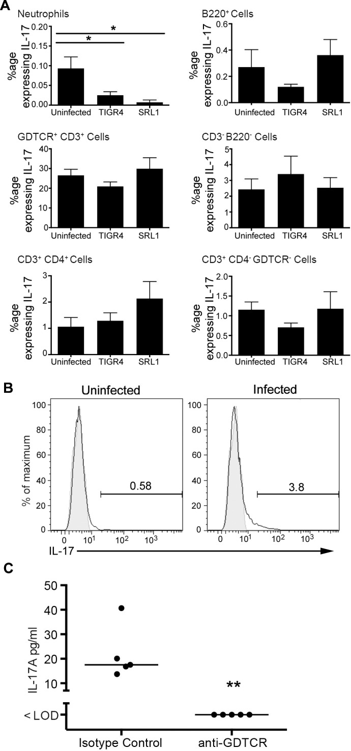
Fig 8. Cellular sources of IL-17A in pneumococcal infection.

(A) Proportion of cells staining positive for IL-17 following stimulation with PMA and ionomycin in the presence of Brefeldin 48 hours post infection. Bars are means of 6 determinations; error bars are SEM. Differences between groups were tested by one-way ANOVA and where significant differences from the uninfected group tested by Dunnett’s test. *, p < 0.05, **, p < 0.01. (B) Flow cytometry of In vivo expression of IL-17 by gamma delta T lymphocytes from animals treated with Brefeldin A 6 hours prior to infection and culled 6 hours post infection. Shaded area is isotype control staining. Representative of 2 independent experiments. (C) BALF IL-17A concentration in animals treated with a gamma delta T cell depleting antibody (anti-GDTCR) or isotype control 24 hours prior to infection and culled 12 hours after infection. Each symbol is an individual animal; line is the median. Difference between the groups is significant (Mann Whitney test, p = 0.008).
