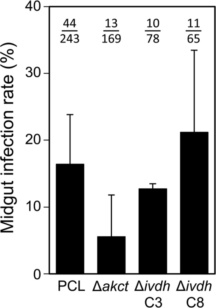
Fig 8. Tsetse fly midgut infection rate with the Δakct, Δivdh-C3 and Δivdh-C8 cell lines.
In this experiment, 50 to 100 Glossina morsitans morsitans teneral males were artificially fed with either the parental (PCL), Δakct, Δivdh-C3 or Δivdh-C8 PCF cell lines in culture medium as previously described (n = 850) [41]. Two weeks after the infective meal, all living flies were dissected to assess the presence of parasites in their entire midgut by microscope examination. Midgut infection rates (in % ±SD) are presented for each strain as the mean of three (mutant cell line) or six (PCL) independent biological replicates (n = 243 for PCL, n = 169 for Δakct, n = 78 for Δivdh-C3, n = 65 for Δivdh-C8).

