Fig. 2.
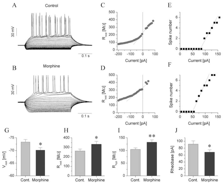
Reduction of the inward rectification in morphine-dependent rats alters the physiological properties of type III neurons. a, b show representative voltage responses of type III neurons from control and dependent rats, respectively. These neurons were stimulated with rectangular current steps of 350 ms duration and increases in the current level at 10 pA increments in the successive cycles. c, d show the input resistance of the two neurons in a and b as a function of the injected current. A strong drop in input resistance at increasingly more negative current levels was associated with activation of the inward-rectifying K+ current. Input-output curves of these neurons are shown in e and f. The rheobase of the neuron from the control rat was ~85 pA, more positive than that of the neuron from the dependent animal. Panels g, h, i, j show the comparison of four physiological parameters of type III neurons. The resting membrane potential of type III neurons from dependent rats was more positive than that from controls (g). The mean input resistance at resting membrane potential (Rrest) was slightly increased in dependent rats (h) and more significantly when measured at −200 pA stimulus levels (Rhyp) (i). The rheobase of type III neurons from dependent animals was significantly reduced relative to control neurons (j).

