
Fig. 4.
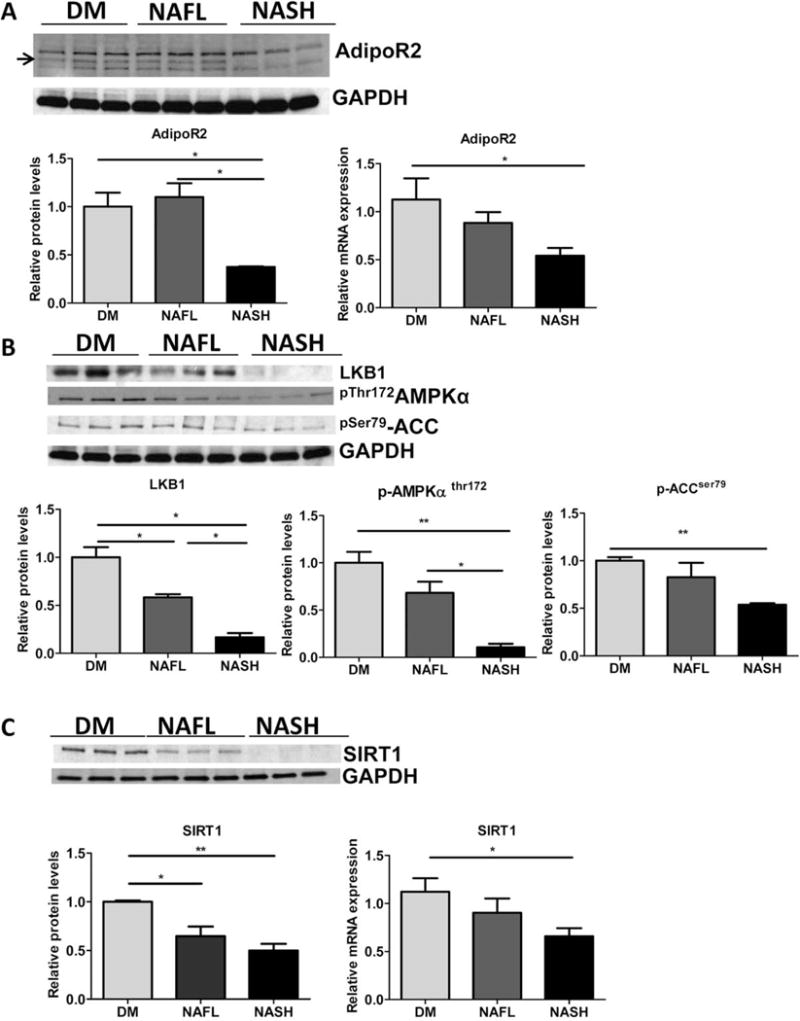
NASH mice experience decrease in hepatic adiponectin-AMPK signaling. Representative western blots (top) and densitometric quantitations of (A) adiponectin receptor-2 and GAPDH protein and relative mRNA expression analysis (below) (B). Representative western blots (top) and densitometric quantitations of LKB1, phosphorylated (Thr 172)-AMPKα protein levels, phosphorylated (Ser 79)-ACCα relative to GAPDH are shown (below). (C). Representative western blots (top), and densitometric quantitations of SIRT1 relative to GAPDH and relative gene expression in DM, NAFL and NASH groups of mice (below). DM mice (n = 5), NAFL (n = 5), and NASH mice (n = 5), *P < 0.05, **P < 0.01 indicates statistical significance between the relevant groups shown.