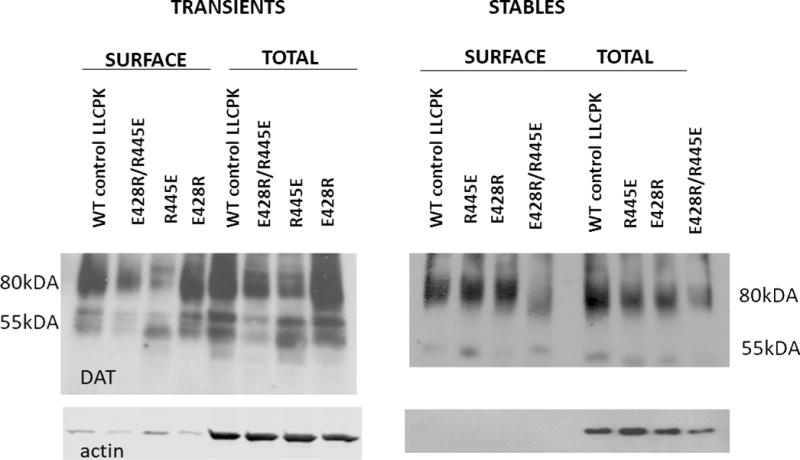
Figure 2.

Immunoblotting studies of mutant hDAT. LLC-PK1 cells transiently (left panel) or stably (right panel) transfected with indicated hDAT constructs were subjected to cell surface biotinylation. Biotinylated proteins (“Surface”) and whole cell lysates (“Total”) were analyzed with antibodies against DAT and actin as indicated. Equal amounts of total lysate protein were loaded for each mutated hDAT as for wild-type (WT).
