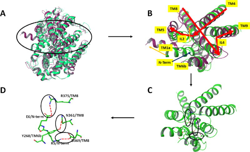
Figure 4.

Comparison via superposition of the outward open (in green, PDB entry 3TT1) and inward open (magenta, PDB entry 3TT3) X-ray structures of LeuT12 viewed from the intracellular side of the proteins. A) Ribbon representation of the full proteins. B) Close-up view of the region encircled in A. Major differences between the 2 protein structures are indicated by orange arrows. Most notably, the N-terminus of the LeuT which is held near the core of the protein through multiple polar interactions in the outward open form (green) is, in the inward-open form, removed from that region (not resolved in the X-ray structure) and TM1a concomitantly swings outward. C) and D) depict the key polar residues comprising the network in the outward open form, in ribbon and stick figures respectively. Molecular graphics were generated with Maestro21.
