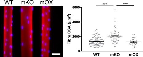
Figure 1.

Typical muscle fibers from WT, mKO, and imOX mice. Single muscle fibers were isolated from the tibialis anterior of age‐matched mKO (SIRT1 muscle‐specific knockout), imOX (SIRT1 muscle‐specific overexpression), and WT (wild‐type) mice. These were then stained for actin (Rhodamine Phalloidin; red) and myonuclei (DAPI, blue). Scale bar: 50 μm. Fiber cross‐sectional areas are shown in the graph (each data point represents one fiber; line and error bars are mean ± SEM)
