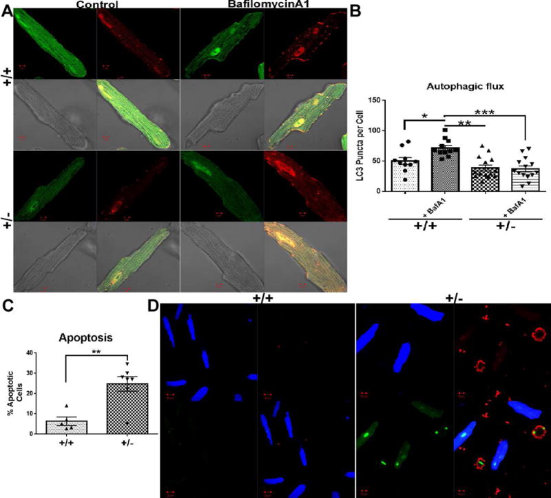
Fig. 3. Effects of BAG3 haplo-insufficiency on autophagy and apoptosis.

(A) Representative confocal images of isolated adult cardiomyocytes from both cBAG3+/+ and cBAG3+/− mice infected with Ad-GFP-RFP-LC3 reporter construct with and without treatment with Bafilomycin A1. (B) Quantification of LC3 puncta per cell. n= at least 9 cells from 3 separate isolations. * p = 0.03, **p = 0.007, ***p <0.0001. (C) Quantification of percent apoptotic cells per field in cBAG3+/+ and cBAG3+/− cardiomyocytes. n= at least 10 fields from 3 separate isolations. p = 0.006. (D) Representative confocal images of cBAG3+/+ and cBAG3+/− stained for phosphatidylserine (red), compromised nuclei (green) and a cytoplasm dye for viable cells (blue).
