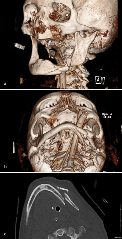
Fig. 1.

( a ) 3D preoperative CT reconstruction of the injury from a three-fourths view. Note the intracapsular head fracture on the left. ( b ) Submental vertex view showing displacement and comminution. ( c ) Axial CT showing comminution. As the maxillary fracture was minimally displaced, the authors believed that any discrepancies would be resolved with an eventual prosthesis.
