Fig. 1.

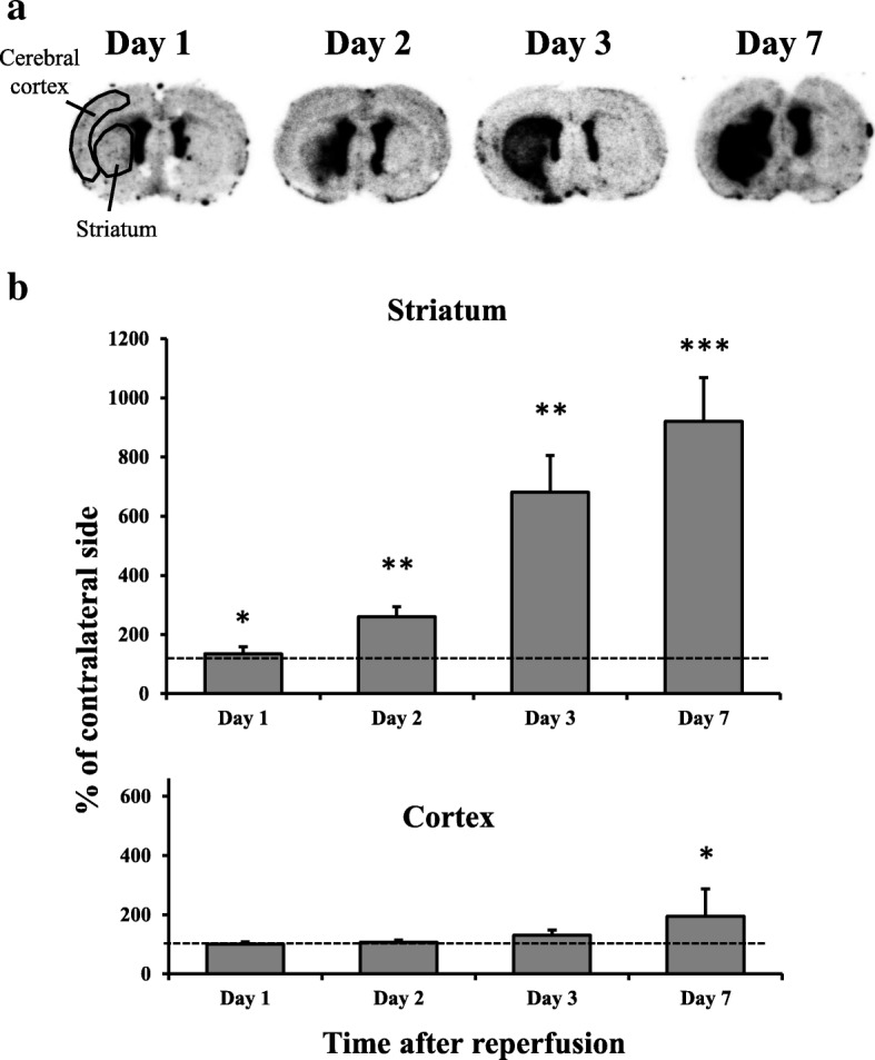
Results of [18F]DPA-714 binding in the brain after mild focal ischemia. a Typical autoradiograms of [18F]DPA-714 binding in the brain after mild focal ischemia. Coronal sections at the level of the striatum were prepared at 1, 2, 3, and 7 days after 20-min middle cerebral artery occlusion (MCAO). The left side of each image is the ipsilateral side. b Temporal changes in [18F]DPA-714 binding in the striatum and cortex after mild focal ischemia. Graphs show % of contralateral side, with values expressed as mean ± SD (day 1: n = 5, day 2: n = 5, day 3: n = 4, day 7: n = 6). Asterisks indicate significant differences compared with the contralateral side, *p < 0.05, **p < 0.01, ***p < 0.001
