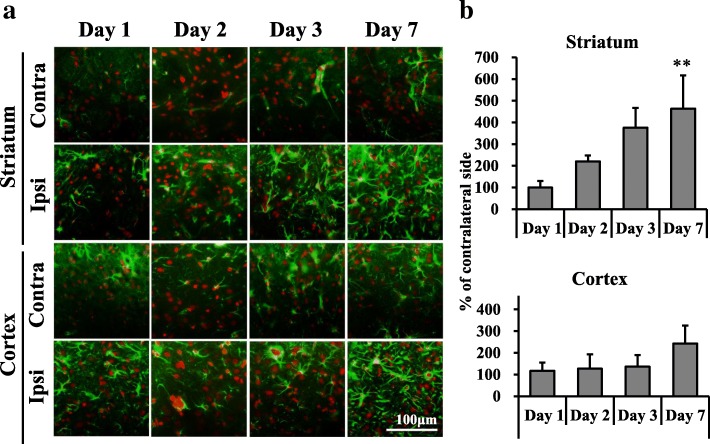
Fig. 6.
Immunostaining with GFAP of the dorsal striatum and cerebral cortex after mild focal ischemia. a Merged immunostaining images were captured with GFAP (green) and PI (red) at 1, 2, 3, and 7 days after 20-min middle cerebral artery occlusion (MCAO). Scale bar, 100 μm. Contra, contralateral side; Ipsi, ipsilateral side. b Graphs show % of the contralateral side, with values expressed as mean ± SD (day 1: n = 3, day 2: n = 3, day 3: n = 3, day 7: n = 3). Asterisks indicate significant differences compared with the contralateral side, **p < 0.01

