
Figure-1.
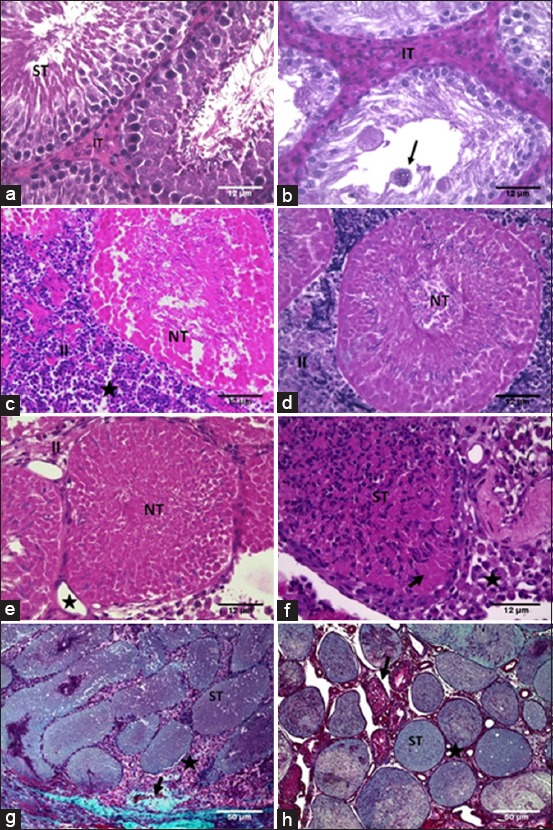
Photomicrographs of testis (hematoxylin-eosin) of adult rats submitted to intratesticular injection of a zinc-based solution and treated with different anti-inflammatory drugs for 7 days and evaluated at 15 and 30 days. (a) Seminiferous tubule (ST) at Stage VII of the seminiferous epithelium cycle and intertubular tissue (IT) in the control group. (b) Animal treated only with dimethyl sulfoxide (DMSO). Note the increased IT and syncytial giant cells (arrow). (c) Necrotic seminiferous tubule (NT) and increased IT (II) with inflammatory infiltrate and cellular debris (star) in an animal treated only with dipyrone. (d) Necrotic seminiferous tubule (NT) and increased IT with inflammatory cells and cellular debris (II) in the celecoxib-treated group. (e) Animal treated with meloxicam, note necrotic seminiferous tubule (NT), increased IT (II) and blood vessels (star). (f) Animal treated with dexamethasone. Note ST within polymorphonuclear leukocyte infiltrates (ST), germ cell necrosis (arrow), and IT with macrophages (star). (g) Animal treated with dipyrone stained using Gomori Trichrome. ST within necrotic germ cells (ST), IT filled by macrophages (star) and fibrosis (arrow). (h) Animal treated only with DMSO stained using Gomori Trichrome. ST within necrotic germ cells (ST) and IT without inflammatory cells (star). Note atrophic ST (arrow).