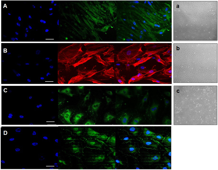
Figure 1.
Representative immunocytochemistry image obtained using a Zeiss LSM700 or LSM 510 confocal microscope. (A) Atrocytes stained with GFAP, (B) pericytes stained with α-actin, (C) endothelial cells stained with CD31 and (D) endothelial cells forming tight junctions (zonula occludens, ZO1) (white arrows). Scale bar = 50 μm. In the right, representative images of the three human primary cell lines – astrocytes (a), pericytes (b) and endothelial cells (c) – cultured independently at early stages (passage 2) are shown. Images were taken from an optical microscope (Nikon Eclipse TS 100) at 20× augmentation.

