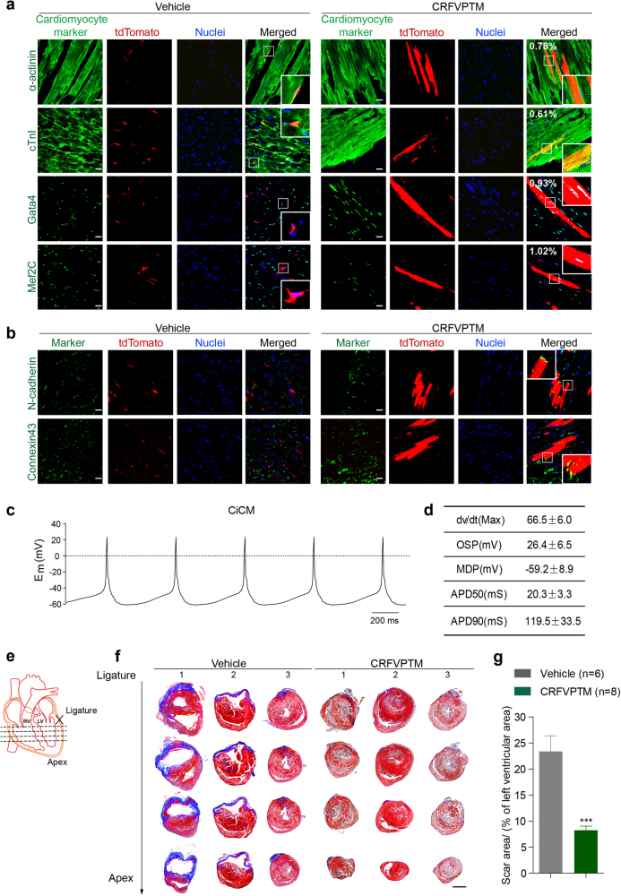
Fig. 1.
Direct reprogramming of adult cardiac fibroblasts into cardiomyocytes in vivo by chemical cocktails. a Eight-week-old Fsp1-cre:R26RtdTomato mice were given CRFVPTM or vehicle once a week for 6 weeks (Supplementary information, Figure S1a, Scheme 5). The animals were then killed and the cryosections of the hearts were stained with antibodies against Mef2C, Gata4, cTnI, and α-actinin. The numbers in the merged images of the drug-treated group represent the percent of tdTomato+ cells expressing various cardiac markers (five sections from each mouse were analyzed, n = 6 for control group and n = 8 for drug-treated group). Nuclei were stained with hoechst. Scale bar, 20 μm. b Immunofluorescence staining of N-cadherin and Connexin 43 in the cryosections of the hearts from Fsp1-cre:R26RtdTomato mice treated with CRFVPTM or vehicle once a week for 6 weeks. Nuclei were stained with hoechst. Scale bar, 20 μm. c Representative action potentials (APs) of the tdTomato+ CiCMs isolated from the hearts of mice treated with CRFVPTM once a week for 6 weeks. APs were recorded in current clamp mode at zero applied current. Em, membrane potential in millivolts. d AP parameters of the tdTomato+ CiCMs, including maximum upstroke velocity (dv/dt Max), over shoot potential (OSP), minimum diastolic potential (MDP), AP durations (APDs) at the level of 50% (APD50) and 90% repolarization (APD90). Data are means ± SEM (n = 7). e Schematic drawing of the positions of the four section levels related to the LAD ligation site. f Representative images (mice 1–3 in both groups) of Masson’s trichrome staining of the heart sections (as presented in e) from mice with LAD ligation and then treated with CRFVPTM or vehicle once a week for 6 weeks (blue areas represent fibrosis, and red areas represent normal tissue). Scale bar, 2 mm. g Quantification of fibrosis area (blue) relative to left ventricular area in heart sections with Masson’s trichrome staining. Four levels from each heart (e) and four slides from each level were measured (a total of 16 sections from each heart. n = 6 for vehicle group and n = 8 for drug-treated group). Data are presented as means ± SEM, ***P < 0.001

