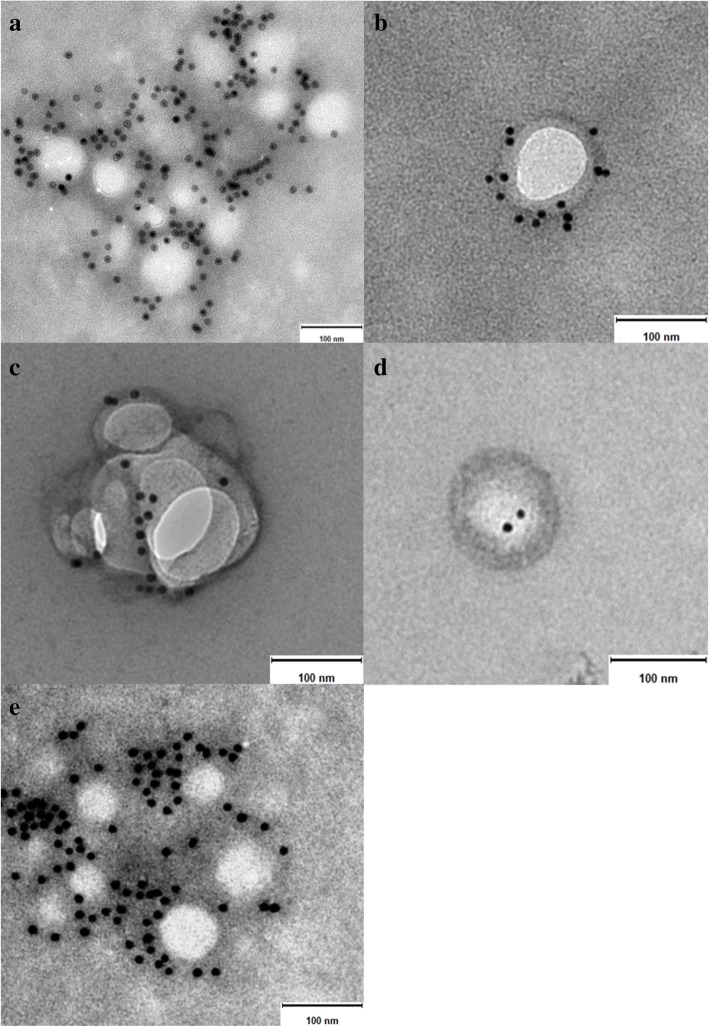
Fig. 3.
Immunoelectron microscopy images of exosomes isolated from samples of canine origin. a and b serum-derived exosomes, c C2 cell line culture-derived exosomes, d and e primary fibroblasts culture-derived exosomes. Note the gold particles bound to the exosome membrane indicating presence of the tetraspanin CD63. Size bar = 100 nm

