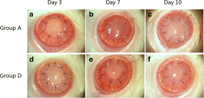
Fig. 2.
Clinical assessment of grafts. Three days post-op, corneal grafts were transparent in Groups A (a) and D (d). Corneal neovascularization began to grow into the cornea from the limbus. At day 7 and when compared to controls (b), both corneal edema and neovascularization of Group D were mitigated (e). By day 10, corneal edema was severe in controls (c). The rejected grafts were opaque and a large number of new vessels had grown into the central portion of the grafts. In MSCs-treated groups (f), the cornea was still transparent with a pupil and new vessels were not near the peripheral portion of the grafts

