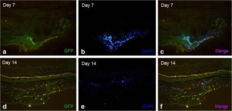
Fig. 7.
MSCs tracking. GFP and DAPI co-labeled MSCs were used in tracking MSCs in subconjuctival sac. GFP-fluorescence (a), DAPI nuclear stain (b, e) and merged (c, f) images are shown. After double injection of MSCs (Day 0, Day 3), there is a large quantity of MSCs can be detected on Day 7 (c). There are still some MSCs can be checked on Day 14 (f)

