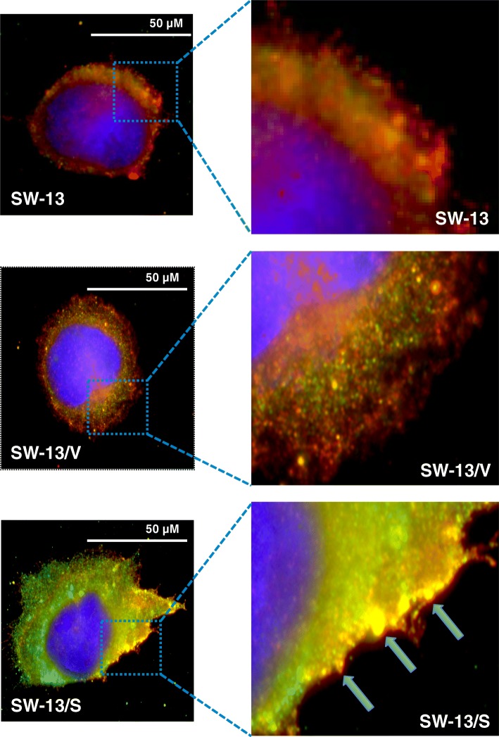
Fig. 7.
Immunofluorescence analysis of SLC12A7 and Ezrin. Cells were grown on glass cover slips in complete medium and after 24 h cells were fixed in cold acetone-methanol (1:1) for 10 min followed by immunofluorescence detection using anti-SLC12A7 and anti-EZR primary antibodies. Cell nuclei fluoresce blue with DAPI, SLC12A7 fluoresces green, and EZRIN fluoresces red. Filled arrows indicate representative areas of co-localization of SLC12A7 with EZR, which fluoresces yellow. SW-13/S cells have heightened expression of SLC12A7 with co-localization with EZR at the cell membrane, especially at the leading edges of the cell when compared to controls (SW-13 and SW-13/V). Immunofluorescence analysis was performed in duplicate

