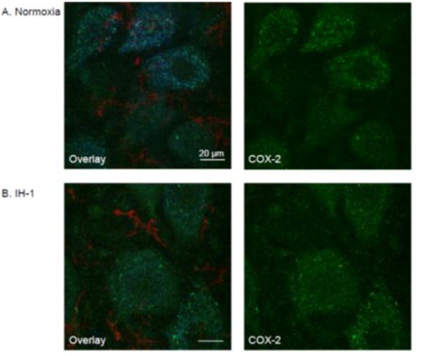
Figure 5.

COX-2 immunofluorescence is similar in phrenic motoneurons after systemic inflammation induced by IH-1 compared to normoxia treated. Confocal images at high magnification (100×) show representative COX-2 staining (green) after normoxia (A, n=6) and IH-1 (B, n=6). COX-2 staining is prevalent in phrenic motoneurons (CtB back-labeled, blue) with only minor expression evident in microglia (CD11b, red) after either treatment.
