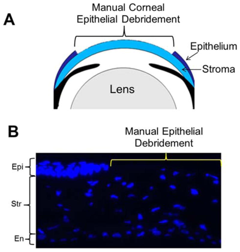
Figure 4.

Corneal debridement. A. Schematic model of the anterior eye showing corneal epithelial debridement method. B. Manual debridement of mouse corneal epithelium. Using Algerbrush II corneal epithelium was carefully scraped off from the cornea. Tissue sections were stained with nuclear stain DAPI (blue). The area demarcated in yellow shows complete removal of the corneal epithelium. The unmarked area shows corneal epithelium and other major layers as labeled. Epi – Epithelium; Str - Stroma; En – Endothelium.
