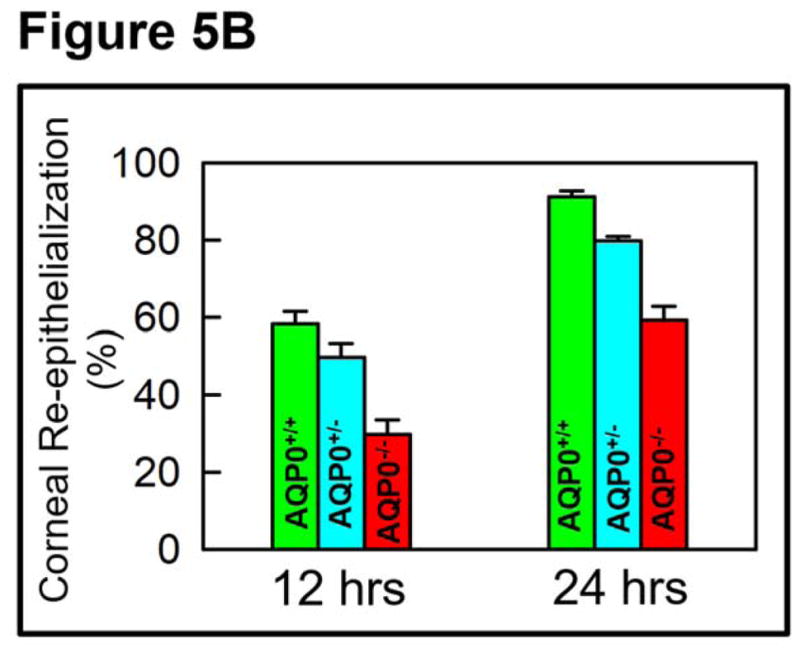
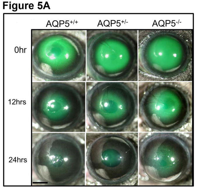
Figure 5.

AQP5-dependent in vivo re-epithelialization after manual corneal epithelial debridement. A. At the center of the cornea, an epithelial debridement of about 2.5 mm diameter was created using Algerbrush II, stained with sodium fluorescein and imaged. Corneal resurfacing was monitored by imaging at regular intervals. Percent of remaining corneal epithelial defects in the wound area was determined by the sodium fluorescein stain remaining at 12 and 24 hrs after wounding. The images are representative of 3 corneas per condition from two independent experiments. Scale bar, 6 mm. AQP5+/+ (wild type); AQP5+/− (heterozygous); AQP5−/− (knockout). B. Rate of wound healing due to corneal re-epithelialization. Wild type (AQP5+/+) expressing AQP5 protein showed higher rate of wound healing (P<0.01) than heterozygous (AQP5+/−) or AQP5 knockout (AQP5−/−).

