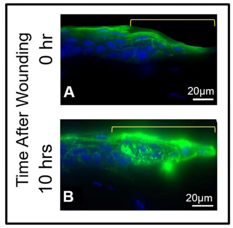
Figure 6.

Immunostaining to show AQP5 expression after corneal epithelial debridement in the epithelial cells and stromal keratocytes from different areas of the cornea of wild type mouse. A. AQP5 expression and localization 0 hr after corneal epithelial debridement, B. AQP5 expression and localization 10 hrs after corneal epithelial debridement. The area marked in yellow on A and B indicates the wound area of the central cornea showing regenerating epithelial cells.
