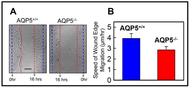
Figure 7.
Ex vivo wound healing model to study mouse corneal epithelial cell migration. A. Corneal epithelial cells were isolated, cultured as monolayers on fibronectin-coated cell culture dishes, scratch-wounded, and repair monitored at regular intervals under a light microscope. Wild type (AQP5+/+) showed faster wound closure than AQP5 knockout (AQP5−/−) 16 hours after wounding. Blue dashed lines: wound margin immediately after wounding. Red dashed lines: cell migration after 16 hours. B. Speed of cell migration of the wound edges of isolated corneal epithelial cells of wild type (AQP5+/+) and AQP5 knockout (AQP5−/−). Wild type (AQP5+/+) showed significantly (P<0.01) faster cell migration than AQP5 knockout (AQP5−/−). Five random sample areas were selected for quantification. Scale bar = 75μm.

