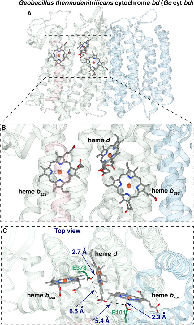
Figure 1. Views of the three hemes in the crystal structure of cyt bd from Geobacillus thermodenitrificans (PDB code: 5IR6 from [12]).

(A) View from the side showing subunit I, containing the hemes, and subunit II (light blue). (B) Closer view from the side of the three hemes. (C) View from the periplasm of the three hemes, also showing the glutamate ligands GtE378 and GtE101, which correspond to EcE445 and EcE99, respectively, from E. coli cyt bd. (Figures made with UCSF package Chimera)
