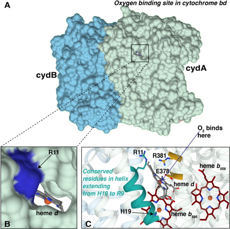
Figure 5. Views of the oxygen binding site in Gt-cyt bd.

(A) The surface representation of subunits I (light green and II (blue) shows the opening leading from the periplasm to the cavity containing heme d. (B) A more detailed view of the opening, with heme d and its Fe (red) visible and with GtR11 (EcR9) indicated on the rim of the opening. (C) A view showing the α-helix that contains both GtR11 (EcR9) and the axial ligand GtH21 (EcH19) to heme b595. This helix runs over the hydrophobic side of heme d. Also shown are GtR381 (EcR448) and GtE378 (EcE445) that form a salt bridge on the hydrophilic side of heme d. (Figure made with Chimera)
