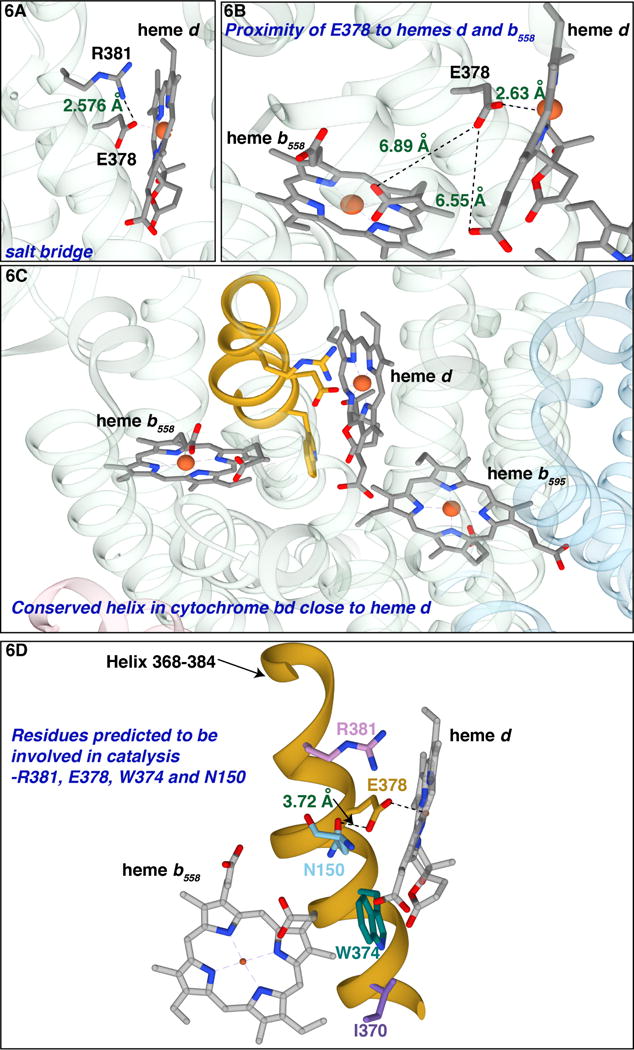
Figure 6. Views of GtE378 and GtR381 in Gt-cyt bd.

(A) The salt bridge between GtE378 (EcE445) and GtR381 (EcR448) at the hydrophilic side of heme d. (B) GtE378 is 6.89 Å from the propionate of heme b558. (C) The conserved α-helix that runs along the hydrophilic side of heme d, 373GWYLA378EVGRQP384W in Gt-cyt bd. (D) The helix is colored in yellow; GtR381, GtE378, GtW374and GtI370 are indicated along with GtN150 (EcN147). (Figure made with Chimera)
