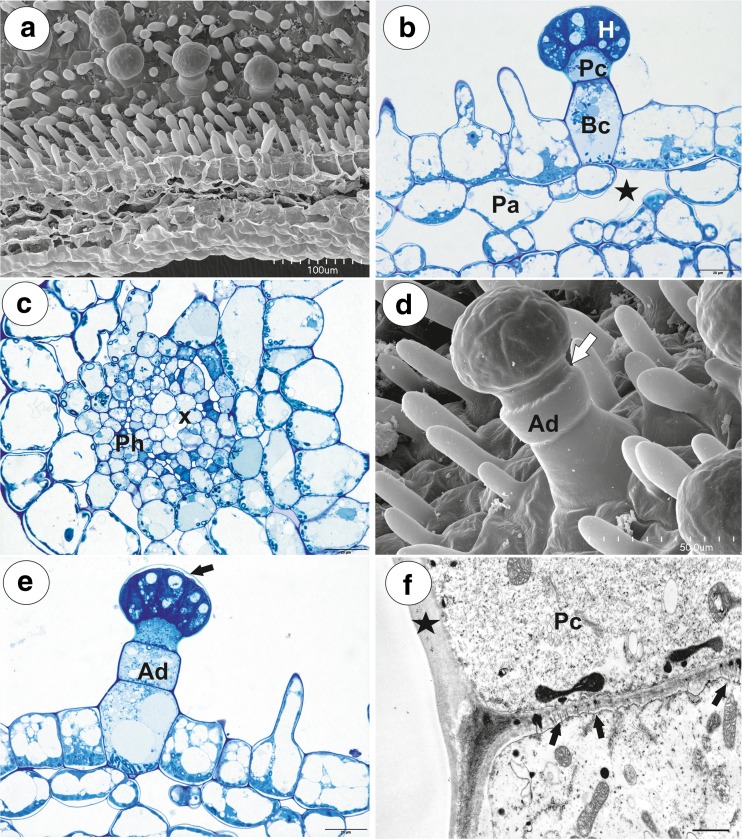
Fig. 2.
Morphology and anatomy of Utricularia vulgaris spur. a Part of the section through the spur with glandular trichomes and papillae; bar = 100 μm. b General structure of the glandular trichome; note that the head cells of the trichomes stain intensely with MB/AII: terminal = head cells (H), pedestal cell (Pc), basal cell (Bc), parenchyma cell (Pa), intercellular spaces (star); bar = 20 μm. c Part of the section through the spur showing vascular bundle: xylem elements (x), phloem (Ph); bar = 20 μm. d, e Trichomes with two-celled stalk consisting of the basal cell and the additional cell (Ad), pedestal cell (white arrow) and separated cuticle from the head cells (black arrow); bar = 50 μm (d) and 20 μm (e). f Part of longitudinal section through basal cell and pedestal cell (Pc), note numerous plasmodesmata (arrows) between these cells; thickened impregnated anticlinal wall of a pedestal cell (star); bar = 1.15 μm

