Fig. 3.
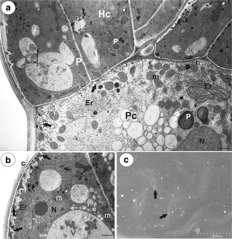
Ultrastructure of a glandular trichome from the spur of Utricularia vulgaris. a Ultrastructure of pedestal (Pc) and terminal cells (Hc); note the well-developed labyrinth wall (arrows) in the pedestal cell, plastids (P), mitochondria (m), endoplasmic reticulum (Er), nucleus (N); bar = 1.2 μm. b Ultrastructure of terminal cells; note the paracrystalline protein inclusion (star) in the nucleus (N), dense cytoplasm with numerous plastids, mitochondria (m). In the vacuoles (V), there is a flocculent electron-dense material. Cell-wall ingrowths (arrows) are on the inner surface of the outer wall; thick cuticle (c); bar = 1.8 μm. c Cuticle of head cells visible in SEM, note cuticular pores (arrows); bar = 5 μm

