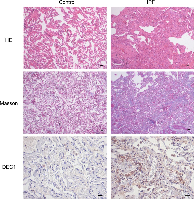
Fig. 5.
The expression of DEC1 in the lung tissue of IPF patients. Normal lungs and fibrosis lungs from patients were taken for hematoxylin-eosin staining, Masson’s trichrome, and immunohistochemistry. a Representative hematoxylin-eosin staining and Masson’s trichrome showed architectural destruction and the presence of dense acellular collagen. b Immunohistochemistry for DEC1 showed the expressions in IPF patients were significantly higher than in controls. n = 5; Scale bar: 200 μm

