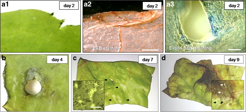
Fig. 1.
Senescence, wound-induced browning and cell death in lettuce fresh-cuts, stored at 4 °C a1 Slight browning at the cut edge (non-labelled tissue). a2 H2O2 production at the area showing initial browning, DAB staining, chlorophyll removed—the tissue appears in brown due to the presence of H2O2. a3 Cell death surrounding an injured site; following Evans Blue staining the dead cells appear in blue (a1–3), day 2. b Tissue browning in area adjacent a site injured with a cork borer, day 4. c Senescing shred on day 7; note sites in which cells have disappeared (arrows); inset shows area with vanished cells. d Senescing shred on day 9; visible is severe browning close to the cut edge, a large necrotic lesion of entirely necrotized tissue and disappearance of cells inside and in vicinity to it (inset, arrows). Scale bars = a2 500 μm, a3 50 μm, c (inset) 100 μm

