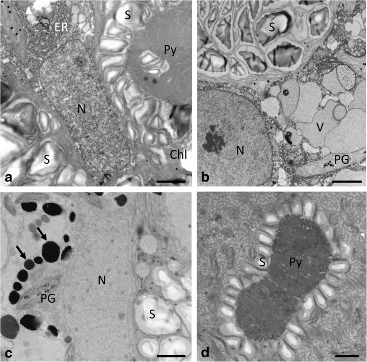
Fig. 7.
Transmission electron micrographs of Zygnema sp. S vegetative cells (a, b) and pre-akinetes (c, d). Cells were exposed either to a PAR+UV-A (PA) or (b–d) to PAR+UV-A+UV-B (PAB). a Central nucleus surrounded by two chloroplasts with prominent pyrenoids, surrounded by numerous starch grains, ER close to the nucleus. b Nucleus with starch-filled chloroplast and individual vacuoles; chloroplast lobes contain plastoglobules. c Central area with nucleus, starch grains in the chloroplast, and electron-dense bodies (arrows) and numerous plastoglobules. d Pyrenoid surrounded by a single layer of starch grains, thylakoid membranes arranged in a cubic structure. Chl chloroplast, ER endoplasmatic reticulum, N nucleus, PG plastoglobules, Py pyrenoid, S starch. Bars 2 μm

