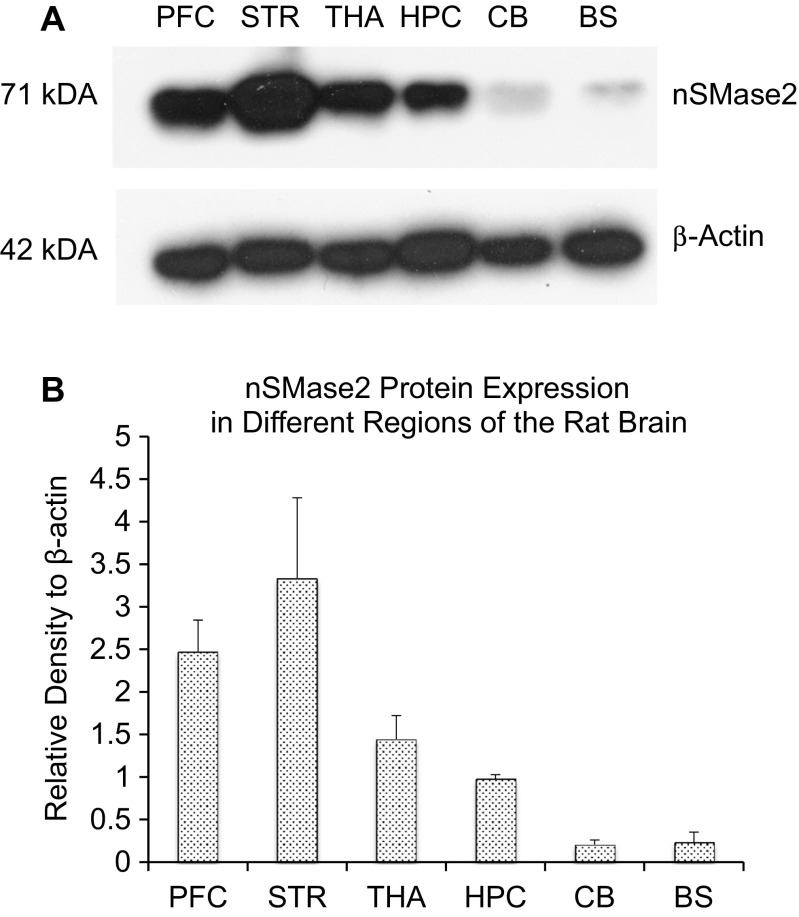
Fig. 3.
a Immunoblot of adult rats in selected parts of the rat brain including the prefrontal cortex (PFC), striatum (STR), thalamus (THA), hippocampus (HPC), cerebellum (CB), and brainstem (BS). The striatum was found to have the highest nSMase2 protein expression, followed by the prefrontal cortex, thalamus, hippocampus, brainstem, and cerebellum. b Densitometric analysis of nSMase2 band intensities, normalized to β-actin. Data represents the mean and standard error from n = 4 Wistar rats. Each bar in the diagram indicates mean + SEM

